.jpg)
JCGenetics offers InSyTe FLECT/CT, a multi-modality preclinical imaging platform that combines the strengths of optical imaging in the form of fluorescence tomography with X-ray computed tomography. The InSyTe FLECT/CT acquires both CT and Industry-first 360° fluorescence imaging for full-angle 3D tomographic imaging.
We also provide top-notch service for current and legacy instruments, as well as application-based support to our customers around the world. TriFoil Imaging is dedicated to helping customers work towards the next scientific breakthroughs in medicine and biology.


ABOUT FLECT
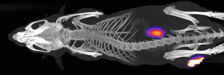
FLECT, or Fluorescence Emission Computed Tomography, is an optical molecular imaging method that uses a full angle tomography data acquisition approach, similar to existing methods such as PET, SPECT, MRI or CT. Unique to FLECT, the optical system is mounted on a gantry that rotates around the animal, allowing full 360° in vivo acquisition of fluorescence emission. This approach is superior in accuracy and sensitivity compared to raster scan approaches used by other optical imaging instruments with limited-angle fluorescence tomography capabilities. FLECT is designed for near-infrared (NIR) fluorescence detection for in vivo deep tissue imaging in mice.
.
How it works ?

The InSyTe FLECT/CT is a small animal in vivo imaging platform that combines FLECT with line X-ray computed tomography to provide optical molecular imaging capability with anatomical reference. The FLECT subsystem is equipped with 4 NIR lasers (642 nm, 705 nm, 730 nm, 780 nm) and corresponding NIR fluorescence emission filters. The lasers are directed at the FLECT gantry and excite fluorescence in mice. A ring of 48 photodiode detectors surrounding the mouse collects the emitted fluorescence.
By rotating the gantry around the subject, the region of interest is illuminated from different angles and the emitted fluorescence captured by the surrounding detectors. FLECT data is acquired in 1 mm slices, with the animal bed moving axially through the FLECT portion of the gantry over the entire region of interest, resulting in 3D data acquisition. Once acquired, the acquired FLECT data is computationally reconstructed into a volumetric image that can be viewed in 3D.